


INVERSE COLLOIDAL CRYSTAL MEMBRANES FOR HYDROPHOBIC INTERACTION MEMBRANE CHROMATOGRAPHY
From a Uniform Pore Structure to Higher IgG Binding Capacity than Commercial PVDF Membranes
Membrane chromatography shortens diffusion distances and operates at lower pressure drops than packed-bed columns. A common limitation is the low surface area and ligand density of membranes, which can restrict binding capacity. This study addresses the problem through support architecture: a membrane with uniform pores, high porosity, and a three-dimensionally interconnected pore network was fabricated and then evaluated for purification of a monoclonal IgG4 antibody by hydrophobic interaction membrane chromatography.
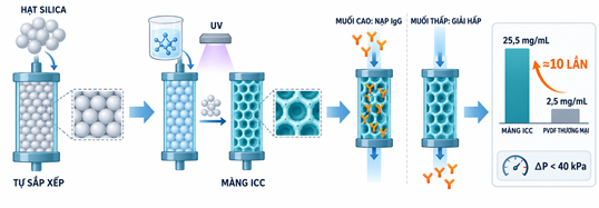
Graphical abstract. Fabrication of the ICC membrane from a silica-particle template, IgG loading and elution in hydrophobic interaction membrane chromatography, and comparison of binding capacity with a commercial PVDF membrane.
Why Membrane Structure Matters
In packed-bed chromatography, solutes must diffuse into the internal pore network of the particles before reaching binding sites. This slows mass transfer, while the column may also experience a high pressure drop when the material deforms or pores become blocked. When a microfiltration membrane is used as a chromatographic support, convective flow carries proteins through the membrane pores and rapidly to the binding surface. However, if the membrane has a broad pore-size distribution or a low surface area, this mass-transfer advantage does not translate into a high adsorption capacity.
In hydrophobic interaction chromatography, the study replaces resin particles with a microfiltration membrane while retaining the same binding mechanism. IgG is loaded in a high-salt buffer and eluted with a low-salt buffer. As the solution passes through the pore network, proteins reach the surface by convection rather than by diffusing deep into packed particles. The membrane therefore needs sufficient binding area while maintaining open flow paths.
Three Key Findings
- The ICC membrane fabricated from 440 nm silica particles had a BET surface area of 27.77 m²/g, compared with 2.21 m²/g for a 0.45 µm commercial PVDF membrane.
- The highest IgG binding capacity reached 25.5 mg per mL of membrane, approximately ten times the 2.5 mg/mL achieved by the commercial PVDF membrane.
- The pressure drop across the membranes remained below 40 kPa in all experiments, and the sharp elution peaks indicated that slow intrapore diffusion had little influence.
Building a Colloidal Crystal Template in a Vertical Chamber
Monodisperse silica particles were prepared by the Stöber method. Mean particle diameters ranged from 300 to 500 nm, with within-batch standard deviations below 5% of the mean. The ethanol suspension was placed in a chamber made from two glass plates and polymer spacers. As ethanol evaporated, capillary flow carried the particles into the membrane-forming region, where they self-assembled into a close-packed structure.
The synthesis used 210 mL ethanol, 17 mL HPLC-grade water, 11 mL ammonium hydroxide, and 11 mL TEOS. The mixture was stirred at room temperature for 4 hours. Particles were centrifuged at 5,000 rpm for 10 minutes and washed three times with 50:50 ethanol/water by volume. Larger particles were obtained by adding 11 mL TEOS and 10 mL water after the first cycle and repeating the 4-hour reaction. The final suspension contained 1-5 wt% particles in absolute ethanol.
The vertical chamber consisted of two 24 × 30 mm cover glasses separated by Mylar strips or microfiltration membranes. Spacer thickness controlled template and ICC-membrane thickness. The microfiltration membrane accelerated ethanol removal and shortened self-assembly compared with Mylar. Depending on particle concentration, the template formed in about one day and was dried at room temperature for 12 hours before monomer infiltration.
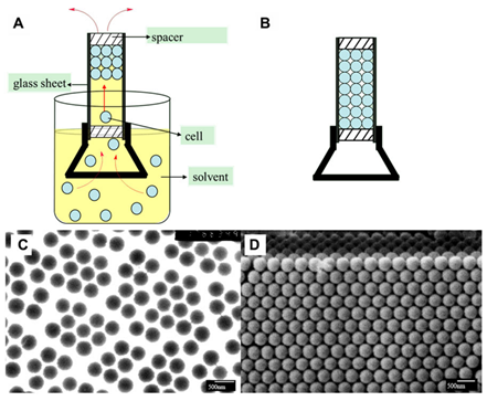
Figure 1. Formation of a colloidal crystal template from 375 nm silica particles: (A) vertical chamber used for self-assembly; (B) template after solvent evaporation; (C) TEM image of the silica particles; and (D) FESEM image of the colloidal crystal template. The figure is reproduced from the original article.
The template formed in approximately one day, depending on the silica-particle concentration. Template thickness, and therefore the thickness of the resulting membrane, was controlled by the spacer thickness. After template formation, the sample was dried at room temperature for 12 hours before monomer infiltration.
TEM images showed that the 375 nm particles were spherical and relatively uniform. FESEM images revealed face-centered close-packed domains, providing the structure needed to generate a repeating, interconnected pore network after silica removal.
Converting the Particle Template into a Three-Dimensional Interconnected Pore Network
The silica template was infiltrated with a mixture of 0.5 g HEMA, 1.5 g HBMA, 0.2 g EGDMA, and 0.03 g BIE photoinitiator. The mixture was polymerized for 15 minutes under a 30 W UV lamp at a wavelength of 254 nm. The silica template was then dissolved in a 10 wt% HF solution, leaving an inverse colloidal crystal (ICC) membrane. 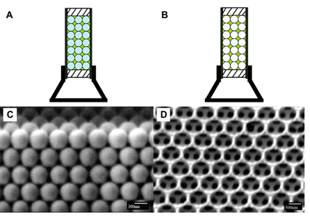
Figure 2. Fabrication of an ICC membrane from a 375 nm silica-particle template: (A) monomer infiltration and photopolymerization; (B) removal of the silica particles to form the membrane; (C) FESEM image of the monomer-infiltrated template after polymerization; and (D) FESEM image of the ICC membrane after template removal. The figure is reproduced from the original article.
What Remains after Template Removal
The spaces originally occupied by the silica particles became pores. The contact points between adjacent particles formed windows connecting the pores, allowing liquid to move through a three-dimensionally interconnected network rather than through isolated cavities. FESEM images showed a relatively uniform structure with only a small number of blocked pores.
The membrane prepared from 440 nm particles had a higher pore density than the membrane prepared from 835 nm particles. Cross-sectional images showed that the pore network extended through the membrane thickness, whereas the commercial PVDF membrane had random pores and a broader pore-size distribution. BET measurements confirmed the difference in surface area: the 440 nm ICC membrane reached 27.77 m²/g, approximately 12.6 times the 2.21 m²/g of the 0.45 µm PVDF membrane.
This design addresses two requirements that often conflict: increasing surface area to provide more protein-contact sites while preserving open flow paths to limit pressure drop.
Comparing the Morphology of ICC and Commercial PVDF Membranes
ICC membranes produced from 440 and 835 nm silica particles were compared with a commercial microfiltration PVDF membrane having a nominal pore size of 0.45 µm. Surface and cross-sectional images showed that the ICC membranes had uniform, clearly interconnected pores, whereas the PVDF membrane had a random pore network with a broader size distribution. 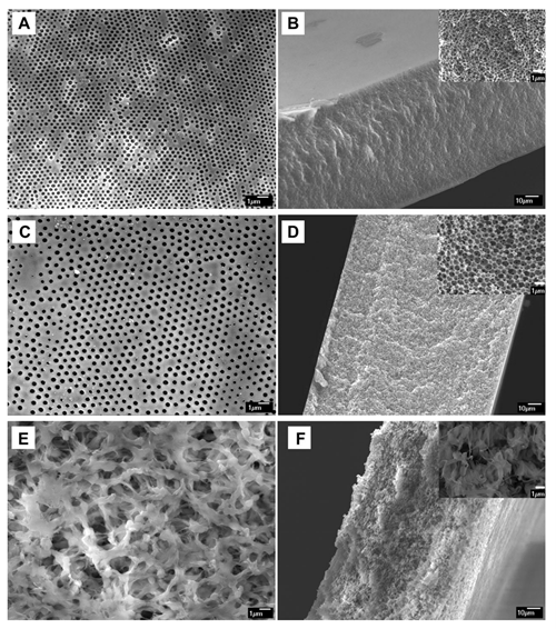
Figure 3. FESEM images: surface (A) and cross-section (B) of the ICC membrane prepared from 440 nm silica particles; surface (C) and cross-section (D) of the ICC membrane prepared from 835 nm particles; and surface (E) and cross-section (F) of the commercial PVDF membrane. The figure is reproduced from the original article.
When pore size decreases in a material of comparable porosity, the number of pores per unit volume increases and the internal surface area also rises. BET measurements showed that the ICC membrane prepared from 440 nm particles had a surface area of 27.77 m²/g, approximately 12.6 times the 2.21 m²/g measured for the 0.45 µm PVDF membrane. This difference explains why the ICC structure can provide more contact area for IgG.
Hydrophobic Interaction Membrane Chromatography Tests with IgG4
The ICC membrane was installed in an ÄKTA FPLC system and tested with a monoclonal IgG4 antibody having a molecular weight of 155 kDa. The base buffer was 20 mM sodium phosphate at pH 7.0, and the high-salt buffer additionally contained 2 M ammonium sulfate. The feed was 1 g/L IgG in high-salt buffer. All solutions were filtered through a 0.22 µm membrane before use.
- Equilibrate the membrane for 30 minutes at 1 mL/min, followed by 2.5 mL of high-salt buffer.
- Load 10 mL of IgG solution at 1 mL/min and wash with 7.5 mL of high-salt buffer.
- Elute IgG with low-salt buffer; the flow rate during washing and elution remains 1 mL/min.
The run comprised four stages: equilibration with 2.5 mL high-salt buffer, loading 10 mL IgG solution, washing with 7.5 mL high-salt buffer without IgG, and elution with low-salt buffer. This separated unretained protein from the fraction recovered after the buffer change. All materials were tested at 1 mL/min for direct comparison of breakthrough position and elution-peak shape. 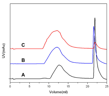
Figure 4. UV absorbance as a function of permeate volume: (A) ICC membrane prepared from 440 nm silica particles; (B) ICC membrane prepared from 835 nm particles; and (C) commercial PVDF membrane. The traces are vertically offset for clarity. The figure is reproduced from the original article.
The breakthrough point shifted to larger volumes in the order PVDF, ICC 835 nm, and ICC 440 nm, consistent with increasing internal surface area as pore size decreases. All three membranes produced sharp elution peaks; the article interprets these shapes as evidence that slow intrapore diffusion did not significantly control the process.
Quantitative Performance and Interpretation of the Results
Table 1. Permeability and Saturated IgG Binding Capacity of the Membranes
|
Membrane |
Permeability |
Binding capacity |
|
ICC from 375 nm particles |
5.0 |
25.5 |
|
ICC from 440 nm particles |
7.0 |
19 |
|
ICC from 835 nm particles |
13.0 |
9.5 |
|
Commercial PVDF (0.45 µm) |
10.3 |
2.5 |
Trade-Off between Permeability and Binding Capacity
Larger-pore membranes showed higher permeability: it increased from 5.0 to 13.0 L·m⁻²·h⁻¹·kPa⁻¹ as template-particle size rose from 375 to 835 nm. IgG binding capacity, however, fell from 25.5 to 9.5 mg/mL. PVDF reached 10.3 L·m⁻²·h⁻¹·kPa⁻¹ but bound only 2.5 mg IgG/mL. The ICC 375 and 440 nm membranes had approximately 10-fold and 7.6-fold higher capacities than PVDF, respectively.
Permeability alone does not describe adsorption performance. The ICC 835 nm membrane had the highest permeability but lower capacity than the two smaller-pore membranes; the 375 nm membrane showed the opposite trend. Pressure drop remained below 40 kPa in every experiment, so the greater surface area did not eliminate the low-pressure advantage of a microfiltration membrane. The appropriate pore size therefore depends on the required balance between binding capacity and throughput.
Conclusion
The results show that membrane-chromatography capacity can be greatly improved by engineering pore geometry, not only surface chemistry. The uniform, interconnected ICC network provides a large surface area while pressure drop remained below 40 kPa. This architecture offers a basis for high-capacity adsorptive membranes in protein purification.
The ICC platform could be functionalized for other chromatographic mechanisms, for example with ion-exchange groups or grafted polymer chains to tune pore size. However, the study evaluated only one model antibody at laboratory scale. Long-term durability, reusability, and performance with complex feeds remain to be established.
Dr. Anh T. Vu - Department of Materials Technology
See more :
- WORKSHOP ON “OPPORTUNITIES AND CHALLENGES FOR WOMEN IN THE ERA OF GREEN TRANSITION”
- BANANA FIBER _ A SUSTAINABLE BIO-BASED ALTERNATIVE TO SYNTHETIC FIBERS
- BANANA FIBER - A SUSTAINABLE BIO-BASED ALTERNATIVE TO SYNTHETIC FIBERS_FCE
- HUIT MATERIALS TECHNOLOGY STUDENTS PARTICIPATE IN INDUSTRIAL STUDY VISITS IN 2026
- ANNOUNCEMENT ON STUDENT CONDUCT SCORES FOR SEMESTER II, ACADEMIC YEAR 2025–2026 – FACULTY OF CHEMICAL TECHNOLOGY